Ghatkopar, Mumbai
+919109108826
+919109108826

WELCOME TO

Dr. Jayesh Pawar

DR. PAWAR is a Consultant Spine Surgeon, practicing spinal surgery in Mumbai. He treats all kinds of spinal problem related to cervical, thoracic and lumbo-sacral areas (neck, upper back& lower back) He has completed his postgraduate Orthopaedic training at the prestigious Krishna Institute of Medical Science, Karad. After completing his training, ...Read More about Dr. Jayesh Pawar
DR. PAWAR is a Consultant Spine Surgeon, practicing spinal surgery in Mumbai. He treats all kinds of spinal problem related to cervical, thoracic and lumbo-sacral areas (neck, upper back& lower back) He has completed his postgraduate Orthopaedic training at the prestigious Krishna Institute of Medical Science, Karad. After completing his training, he was extensively trained in Spine Surgery Fellowship for a period of 6 years in one of the top spinal centres across the globe. He trained with renowned spine surgeons for 4 years at Sancheti Hospital, Pune and Nanavati Super Speciality Hospital, Mumbai. Following that, he was selected on scholarship basis for 1 year training at Macquarie University Hospital, Sydney in Adult and Pediatric Spine Deformity, which is one of the top orthopaedic hospital in Australia. Dr Pawar also did 1 year training at Royal Brisbane And Woman's Hospital, Brisbane in Anterior Spine and Disc Replacement Surgeries, which is one of the largest hospital in Australia. As a Spine Surgeon, he has rich experience of more than 10 years. He is actively involved in research in the field of spine surgery and has publish many articles in international journals. He had also presented his research work at various national and international meetings.
about-cover-bg image
About Dr. Jayesh Pawar

The Best Spine Surgeon in ghatkopar, mumbai

Procedures
Featured Procedures

What We Do

Slip Disc / Sciatica

Slip disc or back disc herniation is most common spinal disorder. Slip disc meaning is protrusion of part of the disc in spinal canal causing pressure on spinal cord and nerves.This leads to inflammation around spinal nerves and will cause Sciatica Causes Primary cause of slip disc is age related degeneration (chronic wear and tear). This causes loss of water content of the disc material leading to loss of it’s shock absorbing capabilities. Patients who are overweight and who have poor lifestyle (wrong posture, long sitting, inactivity) are more prone to develop these degenerative changes. Occasionally, acute injury and accident also can be the cause of disc prolapse. Slip Disc Symptoms: A disc herniation can be totally asymptomatic, especially if the size of disc herniation is small. However, in majority of the cases, the slipped disc causes varying amount of symptoms depending upon size herniated disc, duration of pressure on nerves and inflammation around the nerve. Initially, patients will feel pain at the site of disc prolapse (neck in case of cervical disc prolapse, upper back in case of thoracic disc prolapse and lower back in lumbar disc prolapse). When spinal nerves in the vicinity starts getting affected, pain and tingling-numbness in the extremities (radiculopathy or Sciatica) which may lead to loss of sensation or loss of muscle power in severe cases. In extreme situation with sudden and severe pressure on spinal cord, patient might lose bladder-bowel control also, which requires urgent spinal cord decompression surgery to prevent permanent damage to the spinal cord. A disc prolapse in cervical and thoracic spine can also cause rigidity in the body (spasticity) with loss of balance. Slip Disc Diagnosis: Primary diagnosis of a slip disc is made based on typical clinical history of local pain in spine with nerve related symptoms in the extremities. On physical examination, spine specialist may also find evidence of nerve stretch and compression signs on upper/ lower extremity in the form of loss of sensation and muscle weakness. An X-RAY is usually done as first line of investigation to rule out major bone abnormality. In severe cases which are not responding to medical treatment and physiotherapy, an MRI Scan is done which will show the level and degree of prolapsed disc, and will help spine specialist determine which kind of treatment will be best for the patient. In certain situations an EMG-NCV (Electromyography-Nerve Conduction Velocity) is done to assess nerve function. A CT Scan is done when detailed assessment of bone structure is required or when MRI cannot be done. Treatment: A proper medical treatment, some amount of activity restriction and slip disc exercises (physiotherapy) provides sufficient pain relief to most of the patients to help them live an active life. For the patients who don’t get relief by medical treatment, various options are available. 1. Epidural Injections: For patients who primarily present with radiculopathy (arm/ leg pain) and don’t have emergency symptoms (loss of sensation, weakness, loss of bladder-bowel control), epidural injection can be tried to provide pain relief to the patient. A small amount of anaesthetic medication and steroid are injected next to the prolapsed disc. This reduces inflammation on spinal nerve as well as disc material to provide relief to the patient. If symptoms recur, injection can be repeated 2-3 times. • Microdiscectomy: Patients who do not get adequate relief after medical treatment or epidural injection, or those who have emergency symptoms need to undergo slip disc surgery with removal of protruded disc material. Microdiscectomy is most commonly done slip disc surgery. A small hole is made on the skin and only the part of disc which has come out is removed. There is immediate relief of symptoms and recovery is very quick. • Endoscopic Discectomy: A small tube like endoscope is inserted in the body under local/ general anaesthesia and protruded disc is removed under magnified endoscopic vision. Recovery from this surgery is very quick and this procedure can be done as day-care also. In patients with associated significant back pain/ neck pain, severe disc degeneration (spondylosis), vertebral slip (spondylolisthesis), spine fracture (spondylolysis) and spine deformity (scoliosis/ kyphosis), a spine fusion/ instrumentation might also be advised by your spine doctor along with the removal of slipped disc. The information provided here is for general educational purposes only. For specific advice regarding back pain treatment, please book an appointment with one of our surgeons.

Cervical Myelopathy

Cervical myelopathy is a form of myelopathy that involves compression of the spinal cord in the cervical spine (neck). Your cervical spine contains seven vertebrae (C1 to C7), with six intervertebral discs and eight nerve roots. Causes One common type of cervical myelopathy is cervical spondylotic myelopathy. The term “spondylotic” refers to one of the possible causes of myelopathy — gradual degeneration of the spine that happens as you age. Therefore, cervical spondylotic myelopathy is more common in people 50 and older. The gradual degeneration of the spine often takes the form of cervical spinal stenosis, which is the narrowing of the spinal canal in the neck. Some people are born with a narrow spinal canal (congenital spinal stenosis) and may experience myelopathy sooner than others if further narrowing occurs. Bulging or herniated discs and bone spurs in the neck are other forms of spinal degeneration that can press on the spinal cord and cause myelopathy. Besides the gradual wear and tear of the spine, cervical myelopathy can also be caused by the ossification (hardening) of the ligaments surrounding the spinal cord, such as posterior longitudinal ligament and ligamentum flavum. The ossification of posterior longitudinal ligament (OPLL) is more common. This means that the soft tissue that connects the bones of the spinal column becomes less flexible and slowly turns into bone (ossification). As the ligament becomes thicker, it starts taking up more space and putting pressure on the spinal cord, which leads to myelopathy. The neck portion of the spine is the most common location for OPLL ossification. Other causes of cervical myelopathy may include: • Rheumatoid arthritis of the neck • Whiplash injury or other cervical spine trauma • Spinal infections • Spinal tumors and cancers Symptoms Cervical myelopathy produces two types of symptoms: the ones you may feel in the neck, and the ones appearing elsewhere in the body at or below the compressed area of the spinal cord. The neck symptoms may include: • Neck pain • Stiffness • Reduced range of motion As the disease progresses, one may experience shooting pain that originates in the neck and travels down the spine. Other cervical myelopathy symptoms may include: • Weakness in the arms and hands • Numbness or tingling in the arms and hands • Clumsiness and poor coordination of the hands • Difficulty handling small objects, like pens or coins • Balance issues Diagnosis The earlier cervical myelopathy is diagnosed, the more successful the treatment is expected to be. However, cervical myelopathy symptoms are not unique to this condition and are often mistaken for “normal” signs of aging. To diagnose cervical myelopathy, your doctor may: • Conduct a physical examination and measure your muscle strength and reflexes. • Conduct further tests, including an MRI scan, an X-ray or a CT myelogram of your neck. • Conduct electrical tests to measure how well the nerves in your arms and hands communicate with your brain through the spinal cord. Treatment There are some nonsurgical options for relieving cervical myelopathy symptoms, including physical therapy and a cervical collar brace. However, to eliminate the compression of the spinal cord and prevent worsening of the condition, surgery is often necessary. There are a few surgical procedures your doctor may recommend for treating cervical myelopathy. Widening of the spinal canal (laminoplasty) can be a good motion- sparing option for some patients. Others may benefit from spine decompression surgery with spinal fusion, which is meant to stabilize the spine after herniated discs, bone spurs or ossified ligaments are fully or partially removed. These surgeries can be performed from the back of the neck (posteriorly) or from the front of the neck (anteriorly). Your doctor will recommend a specific surgery approach based on your situation. The information provided here is for general educational purposes only. For specific advice regarding back pain treatment, please book an appointment with our surgeon.

Lumbar Canal Stenosis

Lumbar spinal stenosis is a narrowing of the spinal canal in the lower part of your back. Stenosis can cause pressure on your spinal cord or the nerves that go from your spinal cord to your muscles. Causes The most common cause of spinal stenosis is osteoarthritis. This is the gradual wear and tear that happens to your joints over time. Spinal stenosis is common because osteoarthritis begins to cause changes in most people’s spine by age 50. That's why most people who develop symptoms of spinal stenosis are 50 or older. Some people are also born with a spinal canal that may be narrower than other people's. Besides osteoarthritis, other conditions or things can cause spinal stenosis: • Narrow spinal canal • Injury to the spine • Spinal tumor • Certain bone diseases • Past surgery of the spine • Rheumatoid arthritis • Herniated disk Symptoms Early lumbar spinal stenosis may have no symptoms. In most people, symptoms develop slowly over time. Symptoms may include: • Back pain • Burning pain going into the buttocks and down into the legs (sciatica) • Numbness, tingling, cramping, or weakness in the legs • Loss of sensation in the feet • A weakness in a foot that causes the foot to slap down when walking. This is also called foot drop. • Loss of sexual ability Pressure on nerves in the lumbar region can also cause more serious symptoms known as cauda equina syndrome. If you have any of these symptoms, you need to get medical care right away: • Loss of bowel or bladder control • Severe or increasing numbness between your legs, inner thighs, and back of the legs • Severe pain and weakness that spreads into one or both legs. This makes it hard to walk or get out of a chair. Diagnosis To diagnose lumbar spinal stenosis, your healthcare provider will ask you questions about your symptoms and do a complete physical exam. During the physical exam, your provider will look for signs of spinal stenosis. These include loss of sensation, weakness, and abnormal reflexes. These tests help make a diagnosis: • X-rays of your lumbar spine. These may show bone growths called spurs that push on spinal nerves, a narrowing of the spinal canal, or both. • Imaging tests. A CT scan or MRI scan can give a more detailed look at the spinal canal and nerve structures. • Other studies. You may need a bone scan, a CT taken after injecting dye into the spinal canal (myelogram), nerve conduction studies, and electromyography, an electrical test of muscle activity. Treatment If you have lumbar spinal stenosis, many types of healthcare professionals can help you, such as arthritis specialists, nerve specialists, surgeons, and physical therapists. Treatment can include physical therapy, medicine, and sometimes surgery. Except in emergencies, such as cauda equina syndrome, surgery is often the last resort. • Physical therapy may include exercises to strengthen your back, stomach, and leg muscles. Learning how to do activities safely, using braces to support your back, stretching, and massage may also be helpful. • Medicines may include nonsteroidal anti-inflammatory drugs that relieve pain and swelling, and steroid injections that reduce swelling. Medicines that treat nerve pain or muscle spasm may also be helpful. • Surgery includes removing bone spurs and widening the space between vertebrae (laminectomy). The lower back may also be stabilized by fusing together some of the vertebrae. In some cases, fusion is used along with decompression. Decompression is easing the pressure on the nerves. The information provided here is for general educational purposes only. For specific advice regarding back pain treatment, please book an appointment with our surgeon.

Need Trusted Second Opinion?

Not sure about your diagnosis or treatment plan? We're here to help.

Doctor's Profile Picture

Dr Jayesh Pawar

Dr Jayesh Pawar

MBBS, MS - Orthopaedics, Fellowship in Spine Surgery Orthopedic surgeon
Ghatkopar, Mumbai

Speciality

Orthopedic surgeon


Education

MBBS, MS - Orthopaedics, Fellowship in Spine Surgery Orthopedic surgeon


Experience

10 years


Mobile

9109108826

Memberships

ASSI -Association of Spine Surgeons of India AO Spine , BSS - Bombay Spine Society , BOS - Bombay Orthopaedic Society , GMA - Ghatkopar Medical Association

Registration No

Image Gallery

Pictures That Speak

Testimonials View All
testimonial image

I had a very wonderful experience with Dr Jayesh pawar, he's really a gem of a person.The way he communicates and behave...
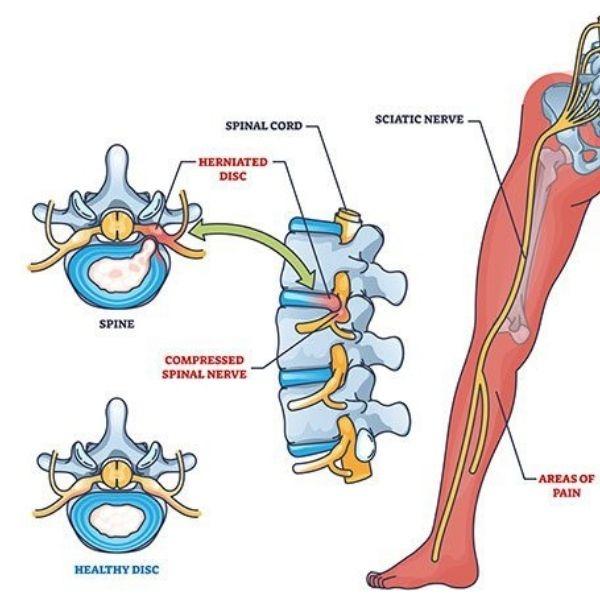
Read More
testimonial image

I visited to doctor Jayesh Pawar with Herniated disc disorder. He consulted me very nicely and suggested me required tre...

Read More
testimonial image

A very humble and caring doctor, always with a smile on the face. Listens patiently, gives clear advice, and treats with...

Read More

footerhc